yourOrthoMD
  • Orthopedic knowledge
    • All Articles
    • All Videos
  • Nutrition
  • Dr. Gorczynski
  • Orthopedic Items

Why is everyone fixated on the meniscus?

2/7/2018

2 Comments

 
With over a million knee arthroscopic surgeries performed in the United States every year, at this point just about everyone has heard about someone having their knee "scoped." This is a quick, low risk procedure that provides relief in the majority of patients.  

Why then am I finding myself spending increasing amounts of time talking patients out of this procedure?

It is important to understand what can be done to a knee with the arthroscope. 
  1. We can photograph normal and abnormal tissue. 
  2. We can remove loose bodies (pieces of broken off cartilage or bone).
  3. We can remove inflamed synovial tissue (joint lining).
  4. We can smooth out unstable articular cartilage flaps. This is known as shaving chondroplasty. 
  5. We can stimulate the bone marrow to induce cartilage healing. This is called micro fracture, and it involves poking small holes in the bone.
  6. We can smooth out/remove a portion of a torn meniscus. 
  7. We can repair some meniscal tears in some people. (The nuances of this are beyond the "scope" of this particular article).
  8. We can reconstruct intra-articular ligaments (the ACL and/or PCL).
  9. We can reinforce weak/inflamed bone by injecting a mineral bone cement (subchondroplasty).

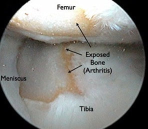
It is crucial to understand one very important thing that can NOT be done with the arthroscope. 

We can not "remove," "clean-up," or otherwise fix arthritis. 

Arthritis involves the permanent loss of articular cartilage. This is the cartilage cushion that coats the bone ends as they meet within the joint. Once articular cartilage is gone, it is gone. The solution when symptoms are unacceptable is joint replacement. Not arthroscopy. 

So what does all of this have to do with the meniscus?

Let me describe an extremely common situation I encounter in my practice. A patient presents to my office with chief complaint of "torn meniscus." They have already had an MRI scan done. Indeed, contained within this report there is reference to a "meniscal tear." There is often reference to some degree of chondromalacia, articular cartilage degeneration, exposed bone, bone marrow edema, or subchondral cysts. All of these terms are essentially saying: "osteoarthritis." 

I always look at these MRI images with patients. I describe all of these pertinent findings. I explain that when there is arthritis in the same knee compartment as a meniscal tear, the pain is most likely due to the arthritis. Even if the meniscus is contributing to the pain, removing it will in no way guarantee pain relief. Many times at this point I am then asked, "so can you clean up the meniscal tear?" It is this fixation on the meniscus that I am attempting to correct. It is crucial to understand that surgically treating a degenerative meniscal tear in an arthritic knee is unpredictable at best. A patient anticipating a miraculous cure can easily be disappointed with the result. 
Arthroscopic view of the knee joint showing healthy articular cartilage and meniscus.
Arthroscopic view of a fairly normal knee joint. The meniscus is the flap of tissue to the left of the picture. The articular cartilage on the bottom (tibia) has a small fissure, it is otherwise healthy appearing. If this meniscus was torn, a good result would be anticipated with arthroscopic surgery.
Arthroscopic photo of an arthritic knee.
This is an arthroscopic view of an arthritic knee. Notice the exposed bone. This problem is clearly advanced far beyond a simple meniscal tear, and it can not be fixed with the arthroscope. Knee replacement would be needed.
The "job" of the meniscus is to help dissipate forces within the joint. It is a shock absorber. By doing this, it helps to prevent arthritis. As our life progresses, this meniscus often wears out as it performs its role. Once the knee becomes arthritic, the only long-term "cure" is knee replacement surgery. 

So what about all those patients that report great relief from knee arthroscopy?

There is definitely a role for knee arthroscopy in an orthopedic practice. With specific regard to our discussion of the meniscus, it is certainly possible to tear the meniscus in the absence of significant arthritis. This is the patient that will predictably do very well with arthroscopic knee surgery. A displaced bucket-handle meniscal tear is a particular tear pattern that results in a "locked knee." A patient usually has difficulty bearing weight, and is unable to fully extend the knee. This condition is an orthopedic urgency. Surgery is required to remove or repair the torn fragment as soon as possible. The urgency is that walking on the displaced meniscus fragment will overload the articular cartilage. This can cause severe irreversible damage to the articular cartilage (arthritis).

​So how do I know who is a good candidate for meniscal surgery? 
​
Standing knee x-rays show joint space narrowing in the setting of osteoarthritis.
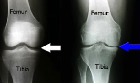
Standing knee x-rays may reveal joint space narrowing. This finding is consistent with arthritis. Arthroscopic surgery to address the meniscal tear in such a situation is likely to be disappointing.
One of the most important studies we do is standing knee x-rays.  Bone partially blocks x-rays whereas cartilage does not. When standing, a healthy knee has a clear separation between the femur and tibia. This separation is known as the joint space. It shows us how much articular cartilage there is. This joint space is not the meniscus, it is articular cartilage. When this joint space becomes narrowed, we know there is some arthritis.  The term "bone-on-bone" arthritis refers to the complete loss of joint space on a standing knee x-ray. This is what severe arthritis looks like.

A good candidate for meniscal surgery is a patient who has been experiencing ongoing pain, swelling, and who has tenderness to palpation over the involved joint line. This is usually made worse by crouching or twisting. Standing x-rays should not reveal much, if any joint space narrowing. An MRI scan should not show articular cartilage loss or bone marrow edema.  In this situation, a knee arthroscopy is a low-risk, high-reward procedure. ​
2 Comments

    Dr. Gorczynski

    Orthopedic Surgeon focused on the entire patient, not just a single joint.

    My YouTube Channel
    ALL ARTICLES

    ALl Videos

    Categories

    All
    Active Range Of Motion
    Adhesive Capsulitis
    Anterior Approach
    Antibiotics
    Arthritis
    Arthroscopic Surgery
    Bursa
    Cartilage
    Cortisone
    Decision For Surgery
    Dental Work
    Dislocation
    Driving
    Extension
    Flexion
    Frozen Shoulder
    Hip Replacement
    Infection
    Inflammation
    Joint Space Narrowing
    Knee Replacement
    Manipulation Under Anesthesia
    Meniscus
    Metal Allergy
    Minimizing Blood Loss
    Numbness
    Partial Knee Replacement
    Passive Range Of Motion
    Patellofemoral
    Platelet Rich Plasma
    Posterior Approach
    Psychology Of Healing
    Quadriceps
    Rehabilitation
    Robotic Joint Replacement
    Rotator Cuff
    Shoulder
    Stiffness
    Stiff Total Knee
    Strengthening
    Stretching
    Tennis Elbow
    Top Doc
    Tourniquet
    Viscoelastic

      Don't Miss Updates from Dr. Gorczynski!

    Sign me up!

    RSS Feed

    Tweets by yourorthoMD

To arrange a private consultation with Dr. Gorczynski:

Email

mailto:chris@yourorthoMD.com
© 2016-2020 Christopher Gorczynski, MD, PLLC.
Photos used under Creative Commons from theglobalpanorama, Tim Evanson, Nicholas_T, Wonderlane
  • Orthopedic knowledge
    • All Articles
    • All Videos
  • Nutrition
  • Dr. Gorczynski
  • Orthopedic Items